Amyloids

Aggregation of protein has been implicated in various neurodegenerative diseases, which mainly includes Alzheimer’s disease (AD), Parkinson’s disease (PD), Multiple system atrophy (MSA) and amyotrophic lateral sclerosis (ALS). The goal of our research is to understand the process of formation of protein aggregates. We study the structural and morphological changes in the protein during aggregation process. We are interested in addressing the correlation between aggregates structure and toxicity. Measurement of the neuronal loss (symptom of neurodegenerative diseases) in mammalian models gives the information about the toxicity of aggregates.

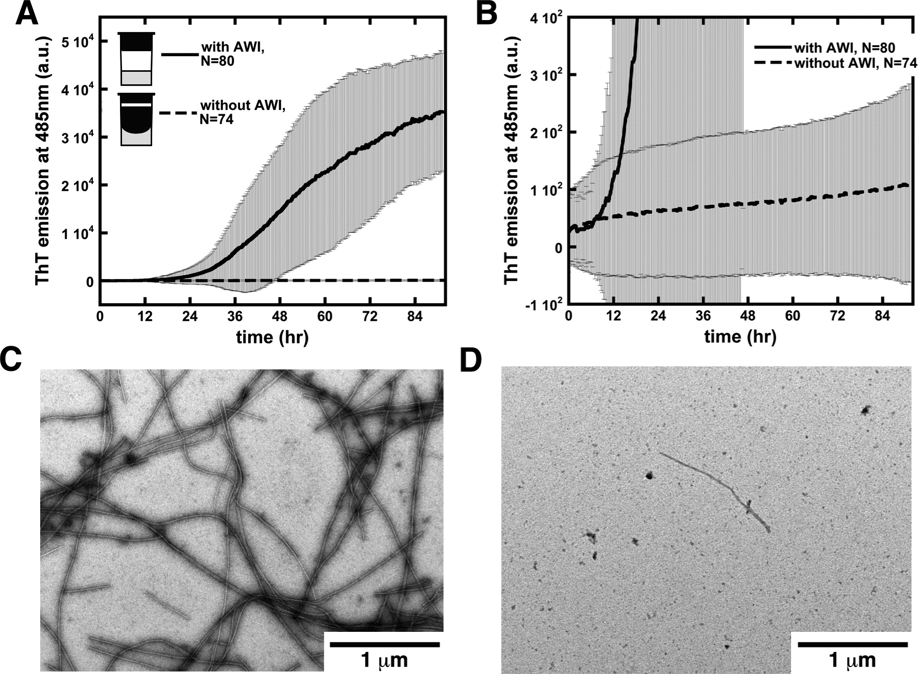
We use several techniques to study the aggregation mechanism, including UV-visible fluorescence spectroscopy, Dynamic light scattering (DLS), Multi-angle light scattering (MALS), Circular dichroism (CD), Electron microscope (EM), Fourier transform infrared spectroscopy (FTIR), Atomic force microscopy (AFM) and Nuclear magnetic resonance (NMR).

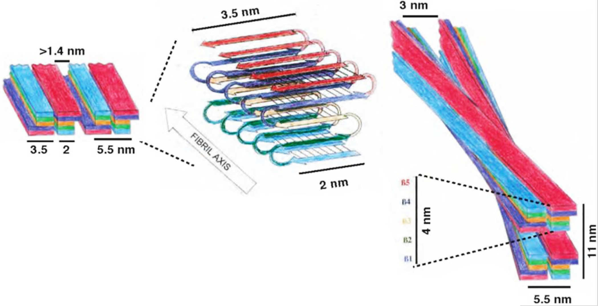
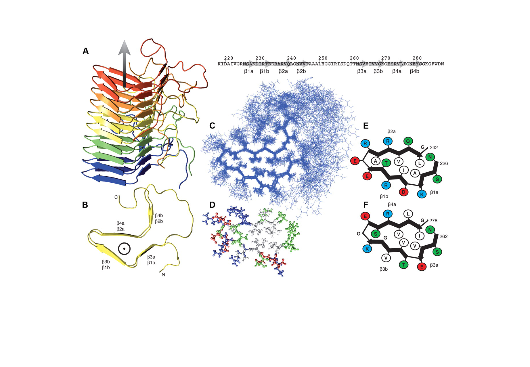
Structure of fibrils:

Enlarged view: sturcture of fibrils
Fig.1. fold of alpha-synuclein fibrils Reference: Vilar M, Chou HT, Lührs T, Maji SK, Riek-Loher D, Verel R, Manning G, Stahlberg H, Riek R. (2008).The fold of alpha-synuclein fibrils. PNAS 105, 8637-42.
Enlarged view: Structure of HET-s fibrils
Fig.2. structure of the HET-s fibrils (Reference: Wasmer C, Lange A, Van Melckebeke H, Siemer AB, Riek R, Meier BH. (2008)Amyloid fibrils of the HET-s(218-289) prion form a beta solenoid with a triangular hydrophobic core. Science, 319, 1523-6. Erratum in Science, 320, 50.)

Aggregation Kinetics:

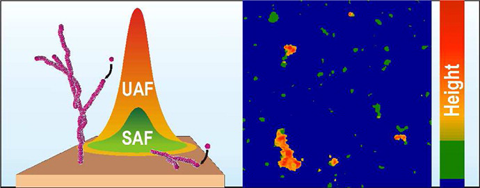
Rate of formation of aggregates, size of aggregates, formation of intermediate aggregates (oligomer and/or proto-fibrils), pathways and morphology of aggregates are found to be dependent on aggregation condition.

Enlarged view: AirWater interface
Fig.3. presence of Air-water interface significantly influences the aggregation kinetics and morphology of aggregates Reference: Campioni, S., Carret, G., Jordens, S., Nicoud, L., Mezzenga, R.,; Riek, R. (2014) The Presence of an Air-Water Interface Affects Formation and Elongation of A-Synuclein Fibrils. J. Am. Chem. Soc. 136, 2866−2875.
Enlarged view: asfadsf
Fig.4. alpha-synuclein fibrils formed by two distinct mechanism Reference: Rabe, M., Soragni, A., Reynolds, NP., Verdes, D., Liverani, E., Riek, R., Seeger, S. (2013) On-Surface Aggregation of α-Synuclein at Nanomolar Concentrations Results in Two Distinct Growth Mechanisms. ASC Chem Neurosci. 4, 408-417.

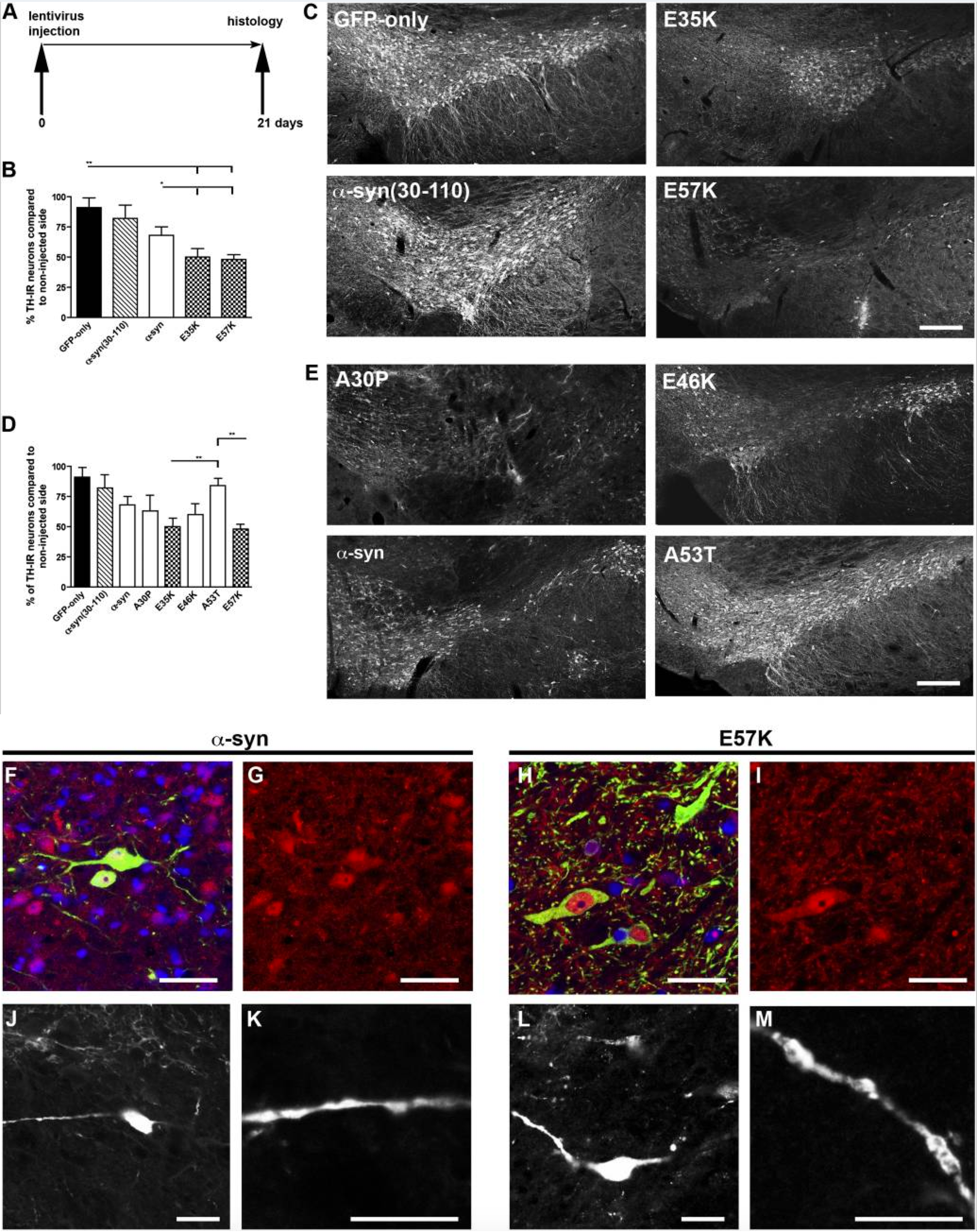
Toxicity of aggregates in mammalian model:

Dopaminergic loss (symptom of Parkinson’s disease) has been monitored in rats. Interestingly variants of alpha synuclein, which form oligomers instead of large aggregates, were found to be the most toxic.

Enlarged view: Toxicity of alpha synuclein
Fig.4. Toxicity of alpha-synuclein Reference: Winner B, Jappelli R, Maji SK, Desplats PA, Boyer L, Aigner S, Hetzer C, Loher T, Vilar M, Campioni S, Tzitzilonis C, Soragni A, Jessberger S, Mira H, Consiglio A, Pham E, Masliah E, Gage FH, Riek R. (2011). In vivo demonstration that α-synuclein oligomers are toxic.
PNAS 108, 4194-9.
JavaScript has been disabled in your browser